43 images of compound microscope with labels
Compound Microscope: Definition, Diagram, Parts, Uses, Working ... - BYJUS A microscope with a high resolution and uses two sets of lenses providing a 2-dimensional image of the sample. The term compound refers to the usage of more than one lens in the microscope. Also, the compound microscope is one of the types of optical microscopes. The other type of optical microscope is a simple microscope. Parts of a Compound Microscope and Their Functions Compound microscope mechanical parts (Microscope Diagram: 2) include base or foot, pillar, arm, inclination joint, stage, clips, diaphragm, body tube, nose piece, coarse adjustment knob and fine adjustment knob. Base: It's the horseshoe-shaped base structure of microscope. All of the other components of the compound microscope are supported ...
Compound Microscope High Resolution Stock Photography and Images - Alamy Find the perfect compound microscope stock photo. Huge collection, amazing choice, 100+ million high quality, affordable RF and RM images. ... Compound Microscope Stock Photos and Images (1,984) compound microscope isolated. Related searches: ... Method of illuminating compound microscope with gas lamp. Labels: C, ...

Images of compound microscope with labels
Labelled Diagram of Compound Microscope - Biology Discussion The below mentioned article provides a labelled diagram of compound microscope. Part # 1. The Stand: The stand is made up of a heavy foot which carries a curved inclinable limb or arm bearing the body tube. The foot is generally horse shoe-shaped structure (Fig. 2) which rests on table top or any other surface on which the microscope in kept. Parts of a microscope with functions and labeled diagram Q. Differentiate between a condenser and an Abbe condenser. Ans. Condensers are lenses that are used to collect and focus light from the illuminator into the specimen. They are found under the stage next to the diaphragm of the microscope. They play a major role in ensuring clear sharp images are produced with a high magnification of 400X and above. Compound Microscope Parts - Labeled Diagram and their Functions - Rs ... Basically, compound microscopes generate magnified images through an aligned pair of the objective lens and the ocular lens. In contrast, "simple microscopes" have only one convex lens and function more like glass magnifiers. [In this figure] Two "antique" microscopes played significant roles in the history of biology.
Images of compound microscope with labels. Compound Microscope Parts, Functions, and Labeled Diagram Compound Microscope Definitions for Labels. Eyepiece (ocular lens) with or without Pointer: The part that is looked through at the top of the compound microscope. Eyepieces typically have a magnification between 5x & 30x. Monocular or Binocular Head: Structural support that holds & connects the eyepieces to the objective lenses. Microscope Types (with labeled diagrams) and Functions A compound microscope: Is used to view samples that are not visible to the naked eye Uses two types of lenses - Objective and ocular lenses Has a higher level of magnification - Typically up to 2000x Is used in hospitals and forensic labs by scientists, biologists and researchers to study micro organisms Compound microscope labeled diagram Compound Microscope - Diagram (Parts labelled), Principle and Uses See: Labeled Diagram showing differences between compound and simple microscope parts Structural Components The three structural components include 1. Head This is the upper part of the microscope that houses the optical parts 2. Arm This part connects the head with the base and provides stability to the microscope. Parts of a Compound Microscope - Labeled (with diagrams) A compound microscope is known as a high-power microscope that enables you to achieve a high level of magnification. Smaller specimens can be thoroughly viewed using a compound microscope. ... Image 3: A compound microscope with a corresponding label of the different parts. imagesource: images.slideplayer.com ... Labels: microsopes Newer Post ...
Compound Microscope Parts, Function, & Diagram - Study.com Learn the compound light microscope's parts and functions by viewing a compound microscope diagram. Also, read about the uses of a compound... Microscope Parts and Functions First, the purpose of a microscope is to magnify a small object or to magnify the fine details of a larger object in order to examine minute specimens that cannot be seen by the naked eye. Here are the important compound microscope parts... Eyepiece: The lens the viewer looks through to see the specimen. Compound microscope Images, Stock Photos & Vectors - Shutterstock Compound microscope images 3,117 compound microscope stock photos, vectors, and illustrations are available royalty-free. See compound microscope stock video clips Image type Orientation Sort by Popular Science College and University Biology Insects and Spiders Jobs/Professions microscope laboratory compound eye optical microscope scientist Next 16 Parts of a Compound Microscope: Diagrams and Video In compound microscopes with two eye pieces there are prisms contained in the body that will also split the beam of light to enable you to view the image through both eye pieces. 2. Arm The arm of the microscope is another structural piece. The arm connects the base of the microscope to the head/body of the microscope.
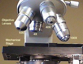
Compound Microscope- Definition, Labeled Diagram, Principle, Parts, Uses In order to ascertain the total magnification when viewing an image with a compound light microscope, take the power of the objective lens which is at 4x, 10x or 40x and multiply it by the power of the eyepiece which is typically 10x. Therefore, a 10x eyepiece used with a 40X objective lens will produce a magnification of 400X. Compound Microscope Labeled Diagram - Quizlet QUESTION. The total magnification of a specimen being viewed with a 10X ocular lens and a 40X objective lens is. 15 answers. QUESTION. a mosquito beats its wings up and down 600 times per second, which you hear as a very annoying 600 Hz sound. if the air outside is 20 C, how far would a sound wave travel between wing beats. 2 answers. 300+ Free Microscope & Laboratory Images - Pixabay 399 Free images of Microscope Related Images: laboratory science bacteria research scientist lab biology chemistry medical Find your perfect microscope image. Free pictures to download and use in your next project. 399 Free images of Microscope / 4‹ › Diagram of a Compound Microscope - Biology Discussion Magnification of the Image of the Object by Compound Microscope 3. Resolution Power 4. Method for Studying Microbes 5. Measurement of the Size of Objects. ... Above positions are repeated using objective lenses and following information's are recorded on an adhesive label. Information recorded on adhesive label is stuck to the base of the ...
Microscope World Blog: Biological Microscope Magnifications
Compound Microscope: Parts of Compound Microscope - BYJUS The parts of the compound microscope can be categorized into: Mechanical parts; Optical parts (A) Mechanical Parts of a Compound Microscope. 1. Foot or base. It is a U-shaped structure and supports the entire weight of the compound microscope. 2. Pillar. It is a vertical projection. This stands by resting on the base and supports the stage. 3. Arm

A Well Labelled Diagram Of A Light Microscope - Top Label Maker
Compound Microscope with labels Stock Vector | Adobe Stock Download Compound Microscope with labels Stock Vector and explore similar vectors at Adobe Stock. Adobe Stock Photos Illustrations Vectors Videos Audio Templates Free Premium Editorial Fonts

Instructions - Microbiology Action - 78 Steps Health
Label Parts Of A Compound Microscope Teaching Resources | TpT This is a set of 3 tiered readings. Students will read a passage about the how to use a compound light microscope. Students will use textual evidence to answer questions and label the different parts of the microscope. It also allows students to gain prior knowledge about the compound microscope. Version A provides the most support for students.
Introduction to the light microscope Flashcards | Easy Notecards
Compound Microscope - Types, Parts, Diagram, Functions and Uses It comes with a wide body and base. Its distinct parts include a condenser, illumination, focus lock, mechanical stage, and a revolving nosepiece which can hold up to five objectives. It usually has a binocular head, which makes long-term observation easy. Image 22: An example of a research compound microscope.
Labeled Compound Microscope - ClipArt Best
Microscope Images Labeled | Virtual Anatomy Lab VAL Microscope Images Labeled | Virtual Anatomy Lab VAL

How to Use a Compound Microscope: 11 Steps (with Pictures)
Compound Microscope stock vector. Illustration of research - Dreamstime Illustration about Clearly labeled vector of modern compound microscope. EPS 8 with no gradients or effects, layers labeled for easy editing. Illustration of research, pathologist, magnify - 23417211
Post a Comment for "43 images of compound microscope with labels"